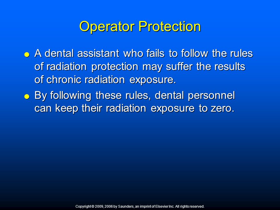
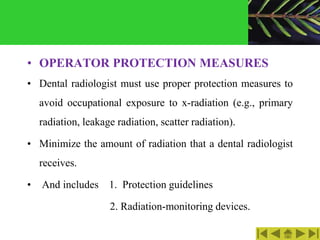
Protection of StaffOperator Use protective devices. Advisable skirt type lead apron to distribute weight.
Foundations Of Radiography Radiographic Equipment And Radiologic Safety Chapter 38 Although This Chapter Covers The Basics Of Conventional Dental Radiography Ppt Download
It is has no weight and travels at the speed of light.

. Identify the types of dental x-ray film holders and devices. ALARA principles are very important to our team as is reducing exposure to excess scatter radiation for our patients. Up-to-date clinical information and compilation of a past imaging record ensures that useless tests are.
Be Aware of All Sources of Radiation Exposure. Justification optimization and dose limitation. Radiation describes any process in which energy emitted by one body travels through a medium or through space ultimately to be absorbed by another body.
Almost complete operator protection can be achieved by a radioprotection cabin or suspended operator protection system that reduces exposure to near background levels including complete protection of the eyes the brain and the axillary region often partially unprotected with lead aprons Figure 7C. Radiation can be classified according to the effects it produces on matter into ionizing and non-ionizing radiations. When shielding is not possible the operator.
One method of reducing radiation exposure in diagnostic radiology is by justifying the examination avoiding repetitions and maintaining complete medical records. 3 Describe the nature of ionizing radiation. This is why certain radioactive materials are.
Explain and Define dental film and apply processing of dental radiographs. Expected levels of radiation from fluoroscopy equipment. When possible operators of radiographic equipment should use barrier protection and barriers should ideally contain a leaded glass window to enable the operator to view the patient during exposure.
When healthcare personnel are in close proximity to the radiation beam they should wear an appropriate lead or lead equivalent apron of sufficient length to shield the upper legs and protect the long bones and bone marrow from increased doses of radiation. The International System of Units SI uses coulombkilogram Ckg in place of roentgen gray Gy instead of rad and sievert Sv rather than rem. This document is designed to provide basic information on radiation mechanisms the dose from various medical radiation sources the magnitude and type of risk as well as answers to commonly asked questions eg radiation and pregnancy.
This variation often occurs due to. Use a Respirator or Face Mask if You are exposed to airborne sources. On completion of this chapter the student will be able to.
Methods of protecting the patient from excess radiation have them wear a lead apron with a tyroid neck color fast speed film the faster the film the less the radiation image receptor for holder devices exposure fraction parell technique. Use a radiation monitoring and stand 6 feet away from an exposure behind a lead barrier or a proper thickness of drywall what are methods used to protect the operator from excess radiation. Therefore we do not allow significant others in the imaging room when we are using ionizing radiation for imaging.
Radiation dose varies widely for different types of examinations and is operator- and facility-dependent as well. Bone and Bone Marrow Protection. One study showed that even for the same type of CT examination the radiation doses varied as much as 13-fold among different institutions and different technologists within the same institution.
As a matter of ease in reading the text is in a question and answer format. Use of lead apron 025 mm lead equivalent radiation dose would be reduced by more than 90 Use ceiling suspended screens lateral shields and table curtains- must for interventional radiology procedures. Describe measures used to protect the operator from excess radiation.
It is a type of electromagnetic wave just like radio waves light waves and x-rays. 5 List the permissible limits of exposure for occupational and. Gamma radiation is a very strong type of electromagnetic wave.
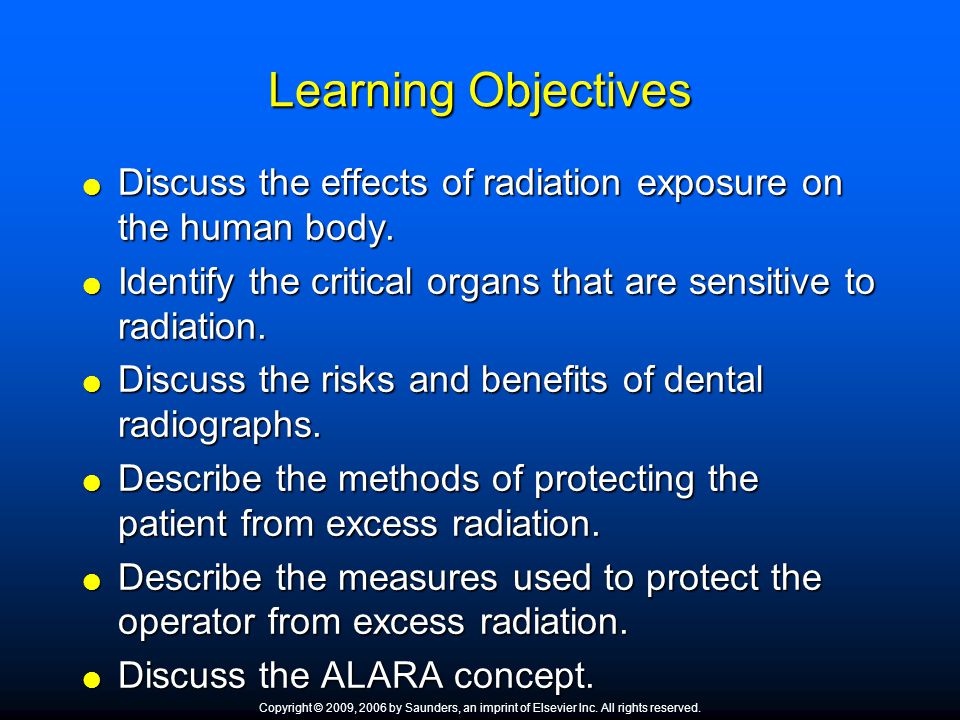
Methods of controlling radiation dose. Characteristics and use of personnel monitoring equipment. Identify the limits of radiation exposure Maximum Permissible Dose or MPD for the operator and the patient.
Describe methods taken to protect the operator from excessive exposure to ionizing radiation including correct position use of barriers and radiation monitoring devices. 2 Describe the units used to measure radiation exposure. Gamma radiation high energy light is a little different.
Know your X-ray history. For radiation protection from x and gamma radiation 1 roentgen Ckg approximately equals 1. The patient must wear an apron that covers from the neck and extends over the lap area to protect from scatter radiation as well as a thyroid collar that is placed around the neck to securely protect the patient.
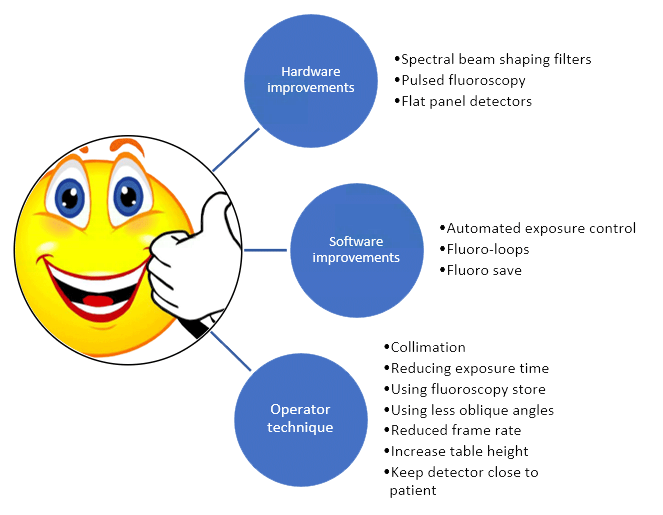
Describe methods of protecting the patient from excess radiation. There are many shielding devices such as caps lead glasses thyroid protectors aprons radiation reducing gloves and so on for radiation safety during C-arm fluoroscopy-guided interventions. Barriers of lead concrete or water provide protection from penetrating radiation such as gamma rays and neutrons.
Significance of radiation dose to include hazards of excessive exposure to radiation biological effects of radiation dose and radiation protection standards. Using digital imaging detectors instead of film further reduces radiation dose. -The operator should never stand in a direct line form the primary beam always stand behind a lead barrier or to the right angles to the beam and never stand closer than 6 feet to the x-ray unit during an exposure unless you are behind a barrier.
This is much faster than alpha and beta radiation. 1 Identify the sources of ionizing radiation. Just as you may keep a list of your medications with you when visiting the doctor keep a list of your imaging records including dental X-rays says Ohlhaber.
Justification involves an appreciation for the benefits and risks of using radiation for procedures or treatments. 15Describe the methods of protecting the operator from excess radiation. Protective measures include the use of barrier shielding occupational radiation exposure limits and personal dosimeters.
Pronounce define and spell the Key Terms. 4 Explain the ways in which ionizing radiation interacts with matter. Measuring Scatter Radiation In Diagnostic X-Rays For Radiation Protection Purposes.
Describe the composition. There are three basic principles of radiation protection. Just as the heat from a fire is less intense the further away you are so the intensity and dose of radiation decreases dramatically as you increase your distance from the source.
Properly Label Sources and keep them Shielded. Ionizing radiation includes cosmic rays X rays and. The rem is a unit that measures the biologic effect of x alpha beta and gamma radiation on humans.
Even though the protective effect is enough for radiation safety no use of the devices cannot protect the physician from radiation. Retrieved from https. We are all exposed to radiation every day from natural sources outer space.
What are methods of protecting the patient from excess radiation. 64 65 They have the added benefit of.
Radiation Safety In The Cath Lab Does It Still Matter Bcs
Foundations Of Radiography Radiographic Equipment And Radiologic Safety Chapter 38 Although This Chapter Covers The Basics Of Conventional Dental Radiography Ppt Download
0 Comments